Structured Training
Workshops and courses on carpal anatomy, palpation, and clinical correlation for healthcare learners.
Evidence-based education and research on the structure, function, and clinical relevance of the wrist and hand.
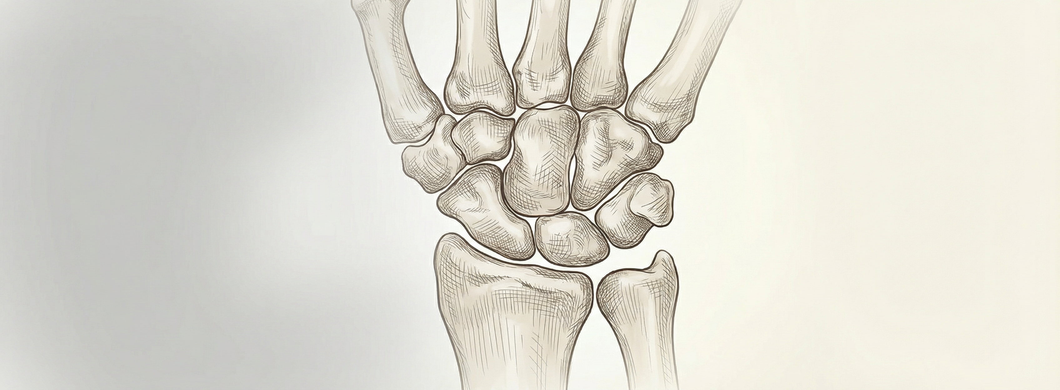
The carpus—eight bones arranged in two rows—forms the mechanical bridge between the forearm and the hand. Understanding their anatomy and biomechanics is essential for accurate diagnosis, rehabilitation, and surgical planning.
Carpanatomy brings together teaching, research, and clinical application. Our materials are designed for students of anatomy, physiotherapy, occupational therapy, and medicine who need a precise, accessible reference grounded in current evidence.
Workshops and courses on carpal anatomy, palpation, and clinical correlation for healthcare learners.
Ongoing work on wrist biomechanics and structural anatomy, with findings applied to teaching and practice.
Guides, references, and materials to support self-directed study and curriculum integration.
The carpal bones are conventionally divided into proximal and distal rows. The proximal row—scaphoid, lunate, triquetrum, and pisiform—articulates with the radius and ulna; the distal row—trapezium, trapezoid, capitate, and hamate—articulates with the metacarpals. Ligamentous and muscular attachments maintain stability while allowing the mobility required for grasp and fine motor tasks.
Pathology in this region—from fractures and ligament injuries to degenerative change—often presents with pain, instability, or loss of motion. A solid grasp of normal anatomy helps clinicians interpret imaging, plan interventions, and communicate findings clearly.
Browse our resources for detailed notes and references on individual bones and clinical applications.
For questions about our programs, research, or resources, or to discuss collaboration, visit our Contact page.
Contact Us